
03 Nov Tooth Root Tip Retrieval in a Cat Patient
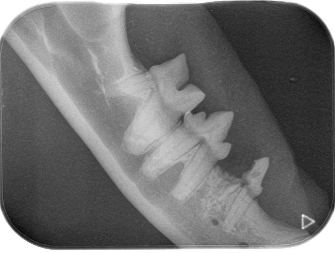
This cat patient, Lola, was referred to me for treatment of tooth resorption disease. The right mandibular premolar in the pre operative dental X-ray below was a very painful tooth and required extraction.
Pre Operative
Teeth with tooth resorption disease can be fragile and surgical extraction can be tricky. It is not uncommon for tooth remnants to be left in after the initial attempt to extract the tooth, as shown in the dental X-ray below. Complete removal of the tooth fragments surrounded by periodontal ligament is necessary to alleviate pain and adequately treat this disease. The root tip fragments in the X-ray below reveals how crucial it was to examine dental radiographs for fragments after extracting these teeth.
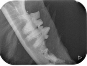
Intra-operative
Here is a zoomed in image of the root tip fragments:
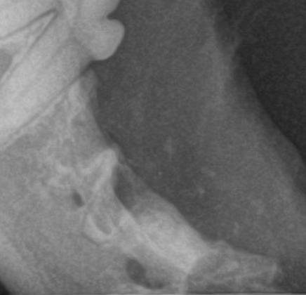
I used a fine headed root tip pic and powerful magnification with surgical operating loops to visualize and retrieve the root fragments. Be sure to use a gentle technique when retrieving root tips, and especially when the root tips are this close to the mandibular canal.
Sometimes I use 1/4 round bur to create a moat around root tips. I do not recommend it in a case such as this, because it is unnecessarily traumatic. I save the 1/4 round bur for more coronally placed root tips.
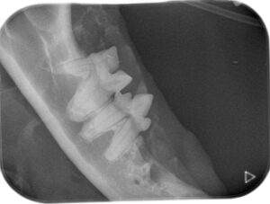
Post operative
It is obvious to the client and us that Lola felt so much better after the procedure. We were happy to send Lola home root tip free.
Best wishes,
Dr. Sharon Startup DVM, DAVDC